Biology (AEES)
Archives of Earth and Environment Sciences
Full Text
Volume 1, Issue 1
Oxidative Stress of Pesticide Residues Leads to Male Infertility
*Corresponding Author: Eman E Elsharkawy, Department of Forensic Medicine and Toxicology, Faculty of Veterinary Medicine, Assuit University, Egypt, E-mail: medicine1971@yahoo.com
doi: /aees.2021.1.102
Citation: Eman E Elsharkawy (2021) Oxidative Stress of Pesticide Residues Leads to Male Infertility. Arch of Earth and Env Sci 1:1-6
Copyright: © 2021 Eman E Elsharkawy. This is an open-access article distributed under the terms of Creative Commons Attribution License, which permits unrestricted use, distribution, and reproduction in any medium, provided the original author and source are credited.
Abstract
Oxidative stress is induced by various pesticides groups. Previous studies suggested that pesticide residues exert oxidative stress as a mechanism of their toxic actions in human or animal’s tissues. The ROS produced by metabolites of pesticides can induce disturbances in the oxidative homeostasis through abnormal redox state and depletion of anti-oxidant. Further, ROS may impair cellular proteins and DNA, and lead to cyto-lethality of the cells via disturbance of signaling pathways and inducing apoptosis pathway. Oxidative stress may be created by residues of and can induce multi reproductive organs dysfunction and male disability.
Keywords: Oxidative Stress; Pesticide Residues; Testicular Damage; Male Infertility.
Definition of the oxidative stress
Oxidative stress creates a disturbance in the systemic activity of reactive oxygen species and in the biological system's capability to deal with the reactive metabolites or to ameliorate the inducing impairment. Imbalance between the normal redox state of cells may induce hazard effects through the induction of peroxides and free radicals that impair the cell contents, including proteins, lipids, and DNA. Oxidative metabolism may inducebasal oxidation, as well as strand breaks in DNA. Reactive oxygen species generated, e.g. O2 − (superoxide radical), OH (hydroxyl radical) and H2O2 (hydrogen peroxide) induced indirect basal damage. In addition, some reactive oxidative species serve as cellular messengers in redox signaling. Though, the normal mechanisms of cellular signaling may be disrupted by oxidative stress [1].
Many pesticide residues have been reported to induce oxidative stress in living systems.
For instance, acetamiprid caused oxidative stress and mitochondrial damage in Leydig cells and increased malondialdehyde and nitric oxide levels in Leydig cells [2]; similarly, thiacloprid also increased oxidative stress [3]. Additionally, carbendazim [4] and chlorpyrifos have been shown to induce oxidative stress by increasing antioxidant enzyme activities and glutathione content and decreasing hydrogen peroxide and lipid peroxidation levels in the hypothalamus, testes and epididymis of treated rats [5]. Furthermore, a mixture of residues (cyhalothrin and imidacloprid) has been shown to induce testicular oxidative stress in adult albino Wistar male rats [6] and to increase testicular malondialdehyde, catalase, superoxide dismutase, and glutathione-Stransferase activities and to reduce testicular glutathione concentrations [7]. Moreover, profenofos has been shown to induce testicular toxicity in mature male rats [8]. Cypermethrin induced oxidative stress and spermatogonial germ cell apoptosis in rats [9]. Similarly, DDT induced testicular oxidative stress in adult rats and increased lipid peroxidation and metallothione in levels, superoxide dismutase catalase activity, and hydrogen peroxide production [10]. Diazinon has been shown to induce oxidative stress by reactive oxygen species, which may be the reason for sperm DNA fragmentation [11]. Dimethoate has been shown to increase the level of lipid peroxidation and to decrease the activities of antioxidant enzymes in the testes of rats [12]. Endosulfan isomers (α endosulfan, β endosulfan and endosulfan sulfate) changed the levels of metabolites involved in energy metabolism and oxidative stress, and these were associated with an imbalance in sex sterol hormone synthesis [13, 14]. Glyphosate, an herbicide residue, decreased glutathione levels and superoxide dismutase activity in the testicular tissue of rats [16]. Similarly, exposure to 2,4-dichlorophenoxyacetic acid has been shown to induce oxidative stress and apoptosis in mouse testes [17, 18]. A mixture of glyphosate and zineb produced severe oxidative stress in testicles by affecting the antioxidant contents [19]. Hexa-chloro-cyclohexane elicited a significant decrease in the activities of cytosolic superoxide dismutase and catalase and ascorbic acid content together with an increase in the levels of lipid peroxidation and hydrogen peroxide [20]. It has been shown that imidacloprid reduced antioxidant activities, increased malondialdehyde content and elevated protein oxidation product levels, with severe histopathological alterations, in rat testes [21]. A mixture of lambda-cyhalothrin and imidacloprid has been shown to increase thiobarbituric acid reactive substance levels, to decrease glutathione levels and to inhibit catalase and superoxide dismutase in Wistar male rats [6].
This evidence shows the potential risk of male infertility. These impairments testicular damage include the following:
1. Mitochondrial damage in Leydig cells caused by acetamiprid [2], clothianidin [22], cypermethrin [23], carbendazim [4], DDVP damage also is caused in Sertoli cells and germ cells of male rats by 2,4-D [24] and carbofuran and in testicular tissues, reducing the diameter of the seminiferous tubules and number of spermatogonia, primary and secondary spermatocytes and spermatids, as caused by chlordane.
2. DNA damage in sperm cells in male mice has recently been shown to be caused by chlorpyrifos [25], glyphosate. [16], α- cypermethrin, and imidacloprid [26].
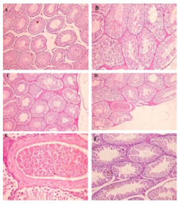
3. Oxidative damage of the testes and testicular tissues in male rats caused by imidacloprid [21] and in the testes of lizards [27]. Moreover, herbicide residues have been shown to create similar effects were observed among farmers in rural area in Malaysia. These herbicides are glyphosate [16], trifluralin [28], and 2,4-D [17, 18]. The exposure to chloropyrifos induces depletion in antioxidant defense systems in the testes This effect may lead to disruption in the functional integrity of cell organelles Figure (1) from pervious ours [29]
4. Necrosis, edema and cellular damage in testicular tissues of rats have been caused by dichlorvos [30, 31]
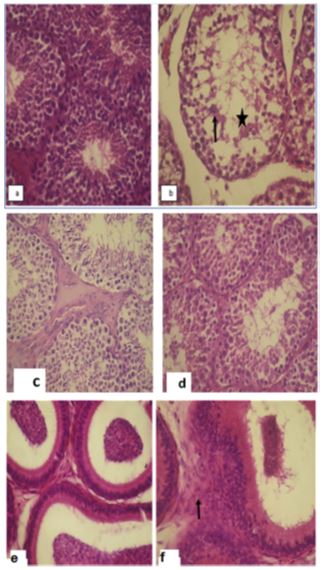
5. Severe seminiferous tubule degeneration in rats has been caused by dimethoate [32], malathion [33] and hexachlorocyclohexane [20, 34]. The prolonged exposure to mancozeb fungicide altered the male rabbit's reproductive abilities and inducing oxidative stress in testicular homogenate. Disruption of the germinal epithelium with vacuolization of Leydig cells and reduced spermatogenic cells Figure 2 from pervious our work [35]
Conclusion
Several pesticide residues can create oxidative stress that may result in dysfunction in testicular cell organelles and altered male reproductive abilities. This evidence shows the potential risk of pesticide residues to induce male infertility.
- El-Nahhal Y (2020) Pesticide residues in honey and their potential reproductive toxicity. Sci. Total Enviro 741: 139953.
- Kong D, Zhang J, Hou X, Zhang S, Tan J, et al. (2017) Acetamiprid inhibits testosterone synthesis by affecting the mitochondrial function and cytoplasmic adenosine triphosphate production in rat Leydig cells. Biol. Reprod. 96: 254-65.
- Kammoun I, Bkhairia I, Ben Abdallah F, Jaballi I, Ktari N, et al, (2017) Potential protective effects of polysaccharide extracted from Ulva lactuca against male reprotoxicity induced by thiacloprid. Arch. Physiol. Biochem. 123: 334-43.
- Sakr SA, Shalaby SY (2014) Carbendazim-induced testicular damage and oxidative stress in albino rats: ameliorative effect of licorice aqueous extract. Toxicol. Ind. Health 30: 259-67.
- Adedara IA, Owoeye O, Ajayi BO, Awogbindin IO, Rocha JBT, et al (2018) Diphenyl diselenide abrogates chlorpyrifos-induced hypothalamic-pituitarytesticular axis impairment in rats. Biochem and Biophys Res Communication 503: 171-6.
- Nantia EA, Kada AS, Manfo FP, Tangu NN, Mbifung KM, et al. (2018) Parastar insecticide induced changes in reproductive parameters and testicular oxidative stress biomarkers inWistarmale rats. Toxicol. Ind. Health 34: 499-506.
- Abdallah FB, Fetoui H, Zribi N, Fakhfakh F, Keskes L (2012) Protective role of caffeic acid on lambda cyhalothrin-induced changes in sperm characteristics and testicular oxidative damage in rats. Toxicol and Ind Health 28: 639-47.
- Moustafa GG, Ibrahim ZS, Hashimoto Y, Alkelch AM, Sakamoto KQ, et al. (2007). Testicular toxicity of profenofos inmaturedmale rats. Arch. Toxicol. 81: 875-81.
- Bhardwaj JK, Kumari P, Saraf P, Yadav AS (2018) Antiapoptotic effects of vitamins C and E against cypermethrin-induced oxidative stress and spermatogonial germ cell apoptosis. J. Biochem. Mol. Toxicol. 32: e22174.
- Marouani N, Hallegue D, Sakly M, Benkhalifa M, Ben Rhouma K (2017) p,p’-DDT induces testicular oxidative stress-induced apoptosis in adult rats. Reprod. Biol. Endocrinol. 15: 40.
- Harchegani AB, Rahmani A, Tahmasbpour E, Kabootaraki HB, Rostami H, et al. (2002) Mechanisms of diazinon effects on impaired spermatogenesis and male infertility. Toxicol. Ind. Health 34: 653-64.
- Jallouli M, El Bini Dhouib I, Dhouib H, Lasram M, Gharbi N, et al. (2016) Disruption of steroidogenesis after dimethoate exposure and efficacy of N-acetylcysteine in rats: an old drug with new approaches. Environ Sci. Poll. Res. Int. 23: 7975-84.
- Yan J, Zhu W, Wang D, Teng M, Yan S, et al. (2019) Different effects of α- endosulfan, β-endosulfan, and endosulfan sulfate on sex hormone levels, metabolic profile and oxidative stress in adult mice testes. Environ. Res. 169: 315-25.
- Aly HA, Khafagy RM (2014) Taurine reverses endosulfan-induced oxidative stress and apoptosis in adult rat testis. Food Chem. Toxicol. 64: 1-9.
- Avdatek F, Birdane YO, Türkmen R, Demirel HH (2018) Ameliorative effect of resveratrol on testicular oxidative stress, spermatological parameters and DNA damage in glyphosate-based herbicide-exposed rats. Andrologia 50: e13036.
- Zhang D, Wu Y, Yuan Y, Liu W, Kuang H, et al. (2017a) Exposure to 2,4 dichlorophenoxyacetic acid induces oxidative stress and apoptosis in mouse testis. Pestic. Biochem. Physiol 141: 18-22.
- Zhang P, Zhu W, Wang D, Yan J, Wang Y, et al. (2017b) Enantioselective effects of metalaxyl enantiomers on breast cancer cells metabolic profiling using HPLC-QT of based metabolomics. Int. J. Mol. Sci. 18: pii: E142.
- Astiz M, Hurtado de Catalfo G, de Alaniz MJ, Marra CA (2012) Exogenous arachidonate restores the dimethoate-induced inhibition of steroidogenesis in rat interstitial cells. Lipids 47: 557-69.
- Samanta L, Sahoo A, Chainy GB (1999) Age-related changes in rat testicular oxidative stress parameters by hexachlorocyclohexane. Arch.Toxicol. 73: 96-107.
- Mahajan L, Verma PK, Raina R, Sood S (2018) Potentiating effect of imidacloprid on arsenic-induced testicular toxicityin Wistar rats. BMC Pharmacol. Toxicol. 19: 48.
- Yanai S, Hirano T, Omotehara T, Takada T, Yoneda N, et al. (2017) Prenatal and early postnatal NOAEL-dose clothianidin exposure leads to a reduction of germ cells in juvenile male mice. J. Vet. Med. Sci. 79: 1196-203.
- Sharma P, Huq AU, Singh R (2014) Cypermethrin-induced reproductive toxicity in the rat is prevented by resveratrol. J. Hum. Reprod. Sci. 7: 99-106.
- Alves MG, Neuhaus-Oliveira A, Moreira PI, Socorro S, Oliveira PF (2013) Exposure to 2,4-dichlorophenoxyacetic acid alters glucose metabolism in immature rat Sertoli cells. Reprod Toxicol. 38: 81-8.
- Kheradmandi R, Jorsaraei SGA, Feizi F, Moghadamnia AA, Neamati N (2019) Protective effect of N-acetyl cysteine on chlorpyrifos-induced testicular toxicity in mice. Int. J. Fertil. Steril. 13: 51-6.
- Zeljezic D, Vinkovic B, Kasuba V, Kopjar N, Milic M, et al. (2017) The effect of insecticides chlorpyrifos, α-cypermethrin and imidacloprid on primary DNA damage, TP 53 and c-Myc structural integrity by comet-FISH assay. Chemosphere 182: 332-8
- Cardone A (2015) Imidacloprid induces morphological and molecular damages on testis of lizard (Podarcis sicula). Ecotoxicol. 24: 94-105.
- Denek Z, Erbil G, Ozbal S, Micili SC, Ozogul C (2016) The effects of resveratrol against trifluralin toxicity in the urinary tract of rats. Toxicol. Ind. Health 32: 106-17.
- Elsharkawy EE, Yahia D and El‐Nisr NA (2014) Chlorpyrifos induced testicular damage in rats: ameliorative effect of glutathione antioxidant. Enviro. Toxicol. 29: 1011-9.
- Dirican EK, Kalender Y (2012) Dichlorvos-induced testicular toxicity in male rats and the protective role of vitamins C and E. Exp. Toxicol. Pathol. 64: 821-30.
- Zeng L, Wang YY, Zhang J, Lin P, Gong XD, et al. (2009) In utero exposure to dichlorvos induces apoptosis of Leydig cells in rats. Zhonghua Nan Ke Xue 15: 1001-6.
- Sayim F (2007) Histopathological effects of dimethoate on testes of rats. Bull. Environ. Contam. Toxicol. 78: 479-84.
- Bustos-Obregon E, Gonzalez-Hormazaba P (2003) Effect of a single dose of malathion on spermatogenesis in mice. Asian J. Androl. 2: 105-7.
- Sujatha R, Chitra KC, Latchoumycandane C, Mathur PP (2001) Effect of lindane on testicular antioxidant system and steroidogenic enzymes in adult rats. Asi. J.of Androl. 3: 135-8.
- Elsharkawy EE, Abd El-Nasser M and Bakheet AA (2019) Mancozeb impaired male fertility in rabbits with trials of glutathione detoxification. Reg. Toxicol. Pharmacol 105: 86-98.